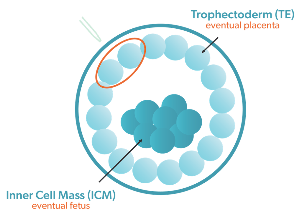
Trophectoderm embryo biopsy is also called blastocyst biopsy
When embryos are biopsied at the blastocyst stage, three to five cells are removed from the trophectoderm of each embryo. This is the part of the embryo that develops into the placenta. The other cell population in a blastocyst, the inner cell mass (ICM), is responsible for creating the fetus, and is not sampled during a trophectoderm embryo biopsy. Your San Antonio fertility specialist will let you know if biopsy for genetic testing of your embryos is recommended.
 What happens in an embryo biopsy?
During the biopsy, precise lasers are used to open a small window in the shell surrounding the embryo, called the zona pellucida. Then, a few cells are removed from the embryo trophectoderm, the part of the embryo that becomes the placenta. These cells are removed with very small instruments connected to a microscope.
Because a blastocyst is made up of 100 or more cells, removing a few cells from the trophectoderm does not impair the embryo’s ability to continue to develop. Embryos develop at different rates, so in our lab, we will perform the trophectoderm embryo biopsy on day 5, 6 or 7 – whenever the embryo develops into a good-quality blastocyst.
The reason a trophectoderm embryo biopsy is done at the blastocyst stage is because it is safer for the embryo and provides more accurate information. When the embryo has only six to eight cells (typical for a day 3 embryo), only one cell can be sampled, and it may not be a true representation of the other cells. If too many cells are taken on day 3, it can affect embryo development to the blastocyst stage, which is required for implantation. Our San Antonio fertility specialists recommend trophectoderm biopsy at the blastocyst stage.
Waiting for results
After the biopsy, the embryos are vitrified, which is a process of quick freezing. The cryopreserved embryo stays at the lab. The cells that were removed are carefully labelled and sent to a genetic testing laboratory. Ovation® Fertility receives the results of the testing, and the patient is informed of the results.
When the patient is ready, a frozen embryo transfer cycle is begun, and one of the tested embryos is transferred to the uterus after preparation with estrogen and progesterone.
Trophectoderm embryo biopsy is most commonly done for preimplantation genetic testing (PGT)
PGT refers to testing done on the embryo prior to an embryo transfer. Most PGT is performed to determine the number of chromosomes present in the cells of the embryo, and to identify whether abnormal numbers of chromosomes are present. Embryos with abnormal numbers of chromosomes are called “aneuploid,” while those with normal chromosome counts are “euploid.”
Embryos should have 46 chromosomes – one copy of each of 22 parental pairs, and either two X chromosomes (female), or an X and a Y chromosome (male). If the embryo has an abnormal number of chromosomes, it will usually fail to implant and the pregnancy test will be negative, or the pregnancy will result in a miscarriage. Embryos with 46 chromosomes are prioritized for transfer.
Sometimes PGT is done to assess whether the embryo has inherited mutated genes leading to childhood illnesses that the parents are carriers for, such as cystic fibrosis, spinal muscular atrophy, or Tay-Sachs disease. This is called PGT-M, or preimplantation genetic testing for monogenic or single-gene disorders.
If a trophectoderm embryo biopsy for genetic testing is recommended, your San Antonio fertility specialist will explain your test options. Contact us to schedule an appointment to learn if you are a good candidate for IVF with PGT.


